
Calculate the peptide's isoelectric point (pI) and net charge before attempting anything else. If aqueous reconstitution fails, escalate through a tiered approach: a volatile pH shift at least two units from the pI, then an organic co-solvent only if that fails. Moving pH away from the pI accelerates certain degradation pathways, so test at small scale first, and check the dilution approach and a peer-reviewed stability review before scaling.
How pH determines peptide net charge, solubility and chemical stability
The isoelectric point (pI) is the pH at which a peptide carries zero net charge. Every ionisable residue, glutamic acid, aspartic acid, lysine, arginine, histidine, and the terminal amine and carboxyl groups, contributes to that balance, and the peptide's solubility tracks its distance from that value almost linearly. At the pI, molecules lack the electrostatic repulsion that keeps them apart in solution, so they aggregate and precipitate. Shift the pH even one unit away, and the accumulating charge starts pushing molecules apart again.
That is the whole logic behind peptide pH adjustment: you're not "dissolving" the peptide chemically, you're manipulating its charge state until electrostatic repulsion overcomes hydrophobic and van der Waals attraction. This is why a sequence that refuses to dissolve in plain water often goes into solution within seconds once the pH moves two units from its pI.
Solubility is only half the story, though. Stability follows a separate, and sometimes conflicting, curve.
Pro Tip: Never assume a peptide that dissolves is a peptide that's stable. Solubility and stability are governed by related but distinct chemistry, and optimising one can quietly compromise the other.
The V-shaped stability curve
Chemical degradation in peptides follows a well-documented V-shaped relationship with pH. Stability is highest across a narrow window, often cited around pH 4.0 to 5.5 for many sequences, and it falls away sharply on either side, according to a review of peptide stabilisation mechanisms. The two arms of that V correspond to different chemistry entirely:
- Acid-catalysed pathways dominate at low pH, chiefly peptide bond hydrolysis, which cleaves the backbone at labile residues (aspartic acid is a common weak point).
- Base-catalysed pathways dominate at high pH, chiefly deamidation of asparagine and glutamine side chains, which converts them to aspartate or isoaspartate and can shift both charge and biological activity.
- Oxidation of methionine, cysteine and tryptophan runs largely independent of pH but often accelerates alongside base-catalysed reactions, compounding the damage; the mechanisms are covered in more detail in a companion guide on peptide oxidation.
- Racemisation at chiral centres, slower than the other pathways but still pH-sensitive, tends to increase toward the alkaline end of the curve.
The stability minimum for a given peptide sits wherever its own sequence dictates, not at a fixed number. A proline-rich sequence behaves differently to one loaded with asparagine residues, and the only way to find the true optimum is a small-scale pH scan rather than a textbook default.
That last point matters more than most protocols admit. Two peptides with similar molecular weights can have stability optima three pH units apart, because the degradation-prone residues sit in different positions and different local environments.
Refrigeration, and even freezing, slows every one of these reactions, but it does not correct a poorly chosen pH. A peptide stored at 4°C in a buffer sitting on the wrong side of its stability curve is still degrading; it's just doing so more slowly. Cold storage buys time, it does not fix chemistry. Researchers who rely on the fridge alone, without first identifying the stability optimum, routinely discover months later that their stock has lost potency despite "proper" cold-chain handling.
Assessing sequence properties that predict solubility
Before touching a buffer or solvent, work out what the sequence is telling you. Three properties do most of the predictive work: net charge at physiological pH, the fraction of hydrophobic residues, and any terminal modification that changes the peptide's charge profile.
- List every ionisable residue. Count aspartate and glutamate as negative contributors, lysine and arginine as positive, and treat histidine as a partial positive contributor below roughly pH 6 given its side-chain pKa near 6.0.
- Check the termini. A free carboxyl C-terminus adds a negative charge above pH 4; an amidated C-terminus (common in synthetic research peptides) removes that charge entirely. A free amine N-terminus adds a positive charge below pH 9; acetylation removes it. These modifications change the sign of the net charge calculation, sometimes by a full unit, so get them right before you calculate anything else.
- Sum the charges at your intended working pH using the standard net-charge method outlined in MerckMillipore's peptide solubility protocol. A result close to zero flags a likely solubility problem regardless of hydrophobicity.
- Estimate the hydrophobic fraction. Tally residues like leucine, isoleucine, valine, phenylalanine, tryptophan and methionine as a percentage of total length.
- Cross-reference the charged-residue ratio. A widely used bench heuristic holds that a sequence needs at least one charged residue for every five amino acids to stay reliably soluble in aqueous buffer, a rule drawn from solvent selection guidance used across peptide protocols.
Pro Tip: Run the net-charge calculation at two pH values, your buffer's target pH and one unit either side, before you commit to a reagent. A sequence that looks borderline at pH 7.4 can look comfortably charged at pH 5.5, and that single calculation can save an entire afternoon of failed dissolution attempts.
Hydrophobic fraction changes the strategy more than most researchers expect. You'll need an organic co-solvent regardless of charge state, because the aggregation driver there is hydrophobic clustering, not electrostatics.
| Sequence signature | Likely issue | Recommended first solvent |
|---|---|---|
| Net charge near zero, low hydrophobicity | Poor solubility from charge neutrality | Volatile pH shift, 2 units from pI |
| Net charge strongly negative (many Asp/Glu, free C-terminus) | Generally soluble at neutral to basic pH | Water or dilute ammonium bicarbonate |
| Net charge strongly positive (many Lys/Arg, free N-terminus) | Generally soluble at acidic pH | Dilute acetic acid |
| High hydrophobic fraction, amidated termini | Hydrophobic aggregation independent of charge | DMSO or acetonitrile co-solvent, then dilute |
| Multiple free cysteines | Disulfide scrambling risk on top of solubility issues | Low concentration, avoid prolonged exposure to basic pH |
Sequences that combine high hydrophobicity with a genuinely amidated, charge-neutral terminus are the hardest cases on the bench, and they're exactly where a hydrophobic solubilisation strategy built around co-solvents earns its place over pH manipulation alone.
Bench protocol for tiered reconstitution
Work through this in order, and resist the urge to jump straight to DMSO because it "usually works." Every tier you skip adds cost, MS incompatibility risk, or unnecessary chemical exposure to the peptide.
Tier 1: aqueous attempt
- Add sterile water or bacteriostatic water directly to the lyophilised peptide, at the wall of the vial rather than directly onto the powder, to avoid disturbing it into a fine aerosol.
- Let it sit for 30 to 60 seconds before swirling gently. Vortexing at this stage often creates foam that traps undissolved particulate and makes visual assessment unreliable.
- Check clarity against a dark background. A faint opalescence can be normal for some longer sequences; visible particulate or persistent turbidity means move to Tier 2.
- If it's close but not quite clear, try brief low-power sonication (30 to 60 seconds in an ice-water bath sonicator) before escalating. Gentle warming to room temperature, never above it for a heat-sensitive peptide, can also help.
Tier 2: volatile pH shift
If aqueous reconstitution fails, shift the pH using a volatile reagent so the peptide can still go on to lyophilisation or mass spectrometry without leaving salt residue.
- For acidic peptides (net negative charge), start with 10 to 100 mM ammonium bicarbonate, prepared fresh, since it loses buffering capacity as it off-gases carbon dioxide over hours.
- For basic peptides (net positive charge), start with 0.1% acetic acid (roughly 17 mM) as the first attempt, moving to more concentrated acetic acid or dilute trifluoroacetic acid only if needed.
- Dissolve the peptide directly into a small volume of the shifted-pH solvent first, then dilute stepwise into your working buffer. Adding the full final volume in one step often causes localised precipitation at the mixing front before the pH has time to equilibrate through the solution.
- Verify pH after full dilution, not just in the concentrated stock, since the working buffer's own buffering capacity can pull the final pH back toward neutral.
This tiered logic, aqueous first, volatile pH shift second, mirrors the standard solvent decision path used across peptide reconstitution protocols, and it's worth following in that exact order rather than skipping ahead.
Tier 3: organic co-solvent
Reserve this tier for hydrophobic sequences that resist both aqueous and pH-shifted attempts.
- Prepare a small concentrated stock in DMSO, DMF, or acetonitrile, whichever your downstream assay tolerates best.
- Dilute that stock into aqueous buffer stepwise, adding the aqueous phase to the organic stock in small increments while agitating gently, rather than the reverse, to avoid a sharp polarity change that reprecipitates the peptide.
- Keep final organic concentration below 10% for most cell-based or enzymatic assays; some tolerate up to 20%, but check your specific assay's published tolerance rather than assuming.
- DMSO is generally the gentlest option for sensitive assays; acetonitrile suits MS-bound samples better since it's more volatile and leaves less residue.
Practical housekeeping matters as much as the chemistry here. Work under sterile technique with filtered reagents, record every volume and pH reading as you go (not retrospectively), and pass the final solution through a 0.22 micron filter if it's headed anywhere near cell culture. A small-volume pH check with a microelectrode before and after each tier gives you a paper trail if something goes wrong three steps later, and it's the single most common step researchers skip when they're in a hurry.
Buffer and solvent selection for downstream compatibility
The buffer that gets your peptide into solution is not automatically the buffer you should keep it in. Downstream use dictates the choice as much as solubility does.
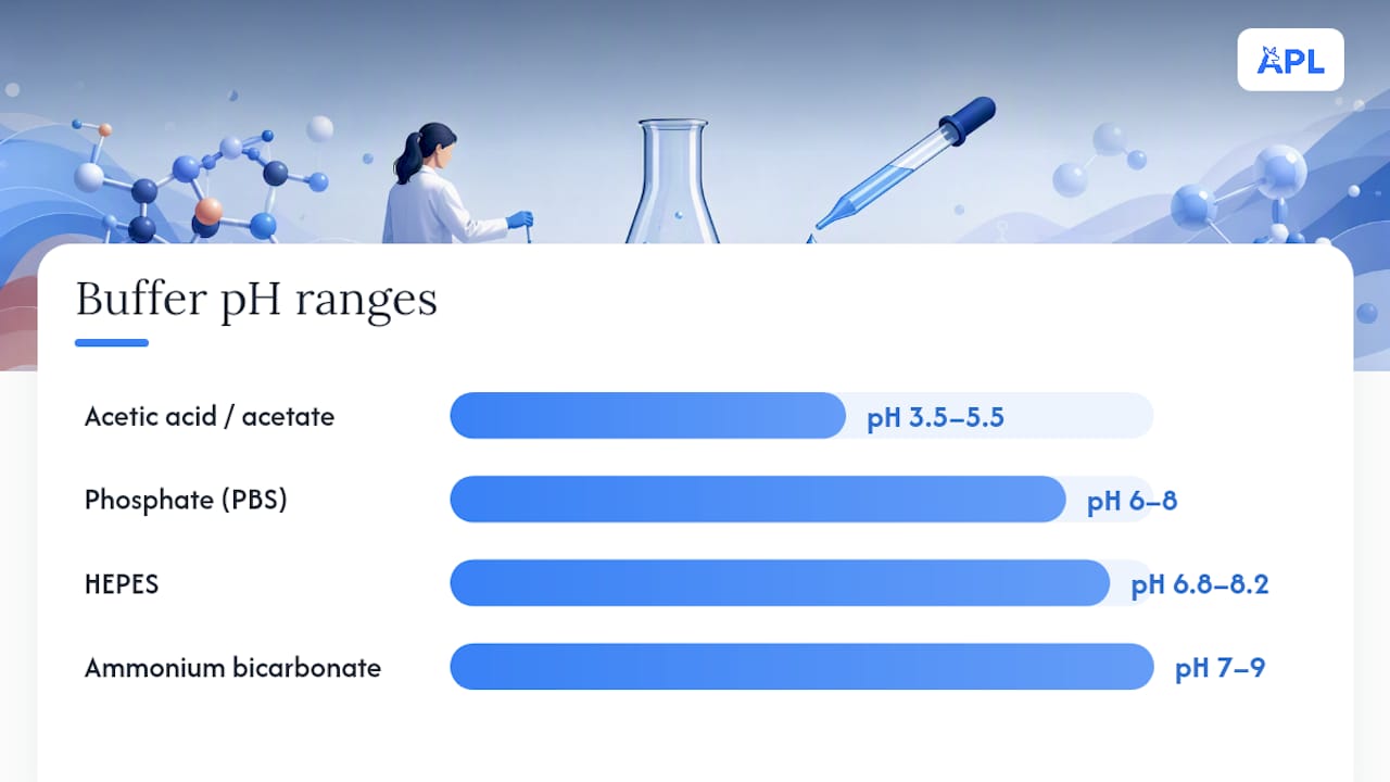
- Ammonium bicarbonate (10 to 100 mM): volatile, MS-compatible, ideal ahead of lyophilisation, but limited buffering range (roughly pH 7 to 9) and it degrades over hours as CO2 escapes.
- Acetic acid / acetate (dilute acetic acid to 100 mM acetate buffer): volatile at low concentration, useful across roughly pH 3.5 to 5.5, good first choice for basic peptides.
- Phosphate buffer (PBS or phosphate buffer, pH 6 to 8): excellent buffering capacity and cell-culture compatibility, but non-volatile, so it leaves salt residue that suppresses ionisation in mass spectrometry and interferes with lyophilisation.
- HEPES (pH 6.8 to 8.2): gentler on biological systems than phosphate, still non-volatile, common in cell-based assay buffers where MS is not part of the workflow.
The MS suppression issue deserves more attention than it usually gets. Non-volatile salts like phosphate and sodium chloride co-elute or co-ionise with the peptide, competing for charge during electrospray ionisation and suppressing your actual signal, sometimes by an order of magnitude. According to protocol discussions among proteomics researchers, volatile buffer systems remain the reliable default wherever MS sits downstream of reconstitution, and switching to a volatile system after the fact, rather than reformulating from scratch, is rarely straightforward once a peptide has already equilibrated in a phosphate buffer.
If your workflow never touches MS or lyophilisation, non-volatile buffers like phosphate or HEPES are entirely reasonable, and often preferable for long-term bench stability given their broader buffering capacity. The decision genuinely comes down to what happens to the sample next, not a universal "always use volatile buffers" rule. When in doubt, or when a sample might move between workflows, default to the lowest ionic strength buffer that still holds your target pH, since it keeps your options open without committing to a particular downstream path too early.
How pH choices affect purification and analytical performance
Reconstitution is only the first place pH matters. It shapes how a peptide behaves through every purification and analytical step that follows, and getting it wrong here can undo everything achieved at the dissolution stage.
- Reversed-phase HPLC typically runs mobile phases at pH 2 to 3 (trifluoroacetic acid or formic acid modified), which fully protonates carboxylic acid groups and gives sharper, more reproducible peaks by suppressing the peptide's ionisation state variability across the column.
- Ion-exchange chromatography works on the opposite principle: choose the running pH specifically to maximise the charge difference between your target peptide and its impurities, since separation resolution depends on that charge gap, not on absolute charge magnitude.
- Ultrafiltration performance is directly pH-sensitive. Peptides near their pI aggregate and foul the membrane, dropping flux and yield, while peptides carrying strong net charge, positive or negative, generally pass through more cleanly and resist membrane binding.
Experimental work on peptide separation confirms that pH shifts measurably change aggregation and separation behaviour during both chromatography and filtration, which means the pH you optimise for reconstitution is not automatically the pH you should run your purification at. A peptide that dissolves beautifully at pH 5 might need to run RP-HPLC at pH 2.5 for peak resolution, then get buffer-exchanged back before storage. Plan for that handoff rather than discovering it mid-run.
Membrane fouling from pH-driven aggregation is worth flagging specifically, since it's a slow, cumulative problem rather than an obvious failure. Flux drops gradually, yield estimates creep down run over run, and researchers often blame the membrane itself before checking whether their working pH sits too close to the peptide's pI.
Measuring pH accurately and storing reconstituted stock
Small-volume pH measurement is where a surprising number of reconstitution "failures" actually originate, not in the chemistry but in the reading. Standard bench electrodes calibrated for 50 to 100 mL samples give unreliable readings in a 20 to 50 microlitre peptide aliquot, and researchers who trust that number without adjusting technique end up chasing a pH shift that was never real.
- Use a microelectrode designed for sub-100 microlitre volumes wherever your working stock is that small.
- Alternatively, dilute a small aliquot into a larger, known volume of pH-neutral water for measurement, then back-calculate, accepting that dilution shifts the reading slightly and building that into your interpretation.
- Recalibrate the electrode at the pH range you're actually measuring, since electrodes calibrated at pH 7 and 10 read imprecisely down at pH 3 to 4.
- Let the reading stabilise fully before recording it; small-volume samples equilibrate more slowly than bulk solution and a premature reading is a common source of error.
Once reconstituted, verify the outcome beyond a visual check. Centrifuge briefly (a benchtop spin at 10,000g for a minute or two is usually enough) and inspect for pellet formation, which flags incomplete dissolution even when the supernatant looks clear. A quick pass through a 0.22 micron filter, followed by a UV absorbance check or an LC injection where instrumentation is on hand, confirms both clarity and that nothing has degraded during the reconstitution itself.
For storage, keep reconstituted stock refrigerated at 2 to 8°C for short-term use, and frozen at negative 20°C or below for anything beyond a few days, protected from light if the sequence includes tryptophan or other light-sensitive residues. Aliquot into single-use volumes before freezing rather than repeatedly freeze-thawing a bulk stock, since freeze-thaw cycles compound both aggregation and chemical degradation risk. Flag any stock that's been sitting outside its optimal pH window, even refrigerated, for re-testing before use in a critical experiment rather than assuming cold storage preserved it fully; how temperature excursions interact with pH-driven degradation is worth understanding before you rely on a stock that's had an uncertain thermal history.
Troubleshooting common pH adjustment problems
| Observation | Likely cause | Corrective action |
|---|---|---|
| Incomplete dissolution after Tier 1 and 2 | Hydrophobic clustering unrelated to charge | Move to Tier 3 organic co-solvent |
| Precipitation on dilution into working buffer | Sharp pH or polarity change at the mixing front | Dilute more slowly, in smaller increments, with gentle agitation |
| Foaming during reconstitution | Vortexing too aggressively, or surfactant-like sequence behaviour | Swirl gently instead of vortexing; let sit before assessing |
| Cloudy solution that clears with time | Slow re-equilibration of pH through the full volume | Wait 10 to 15 minutes and recheck before escalating tiers |
| Unexpected extra LC peaks | Deamidation or hydrolysis from prolonged exposure at a poor pH | Re-test a fresh vial; review storage pH and time since reconstitution |
| Pellet after centrifugation despite clear supernatant | Partial aggregation, not full dissolution | Repeat Tier 2 with a slightly larger pH shift from pI |
Distinguishing reversible aggregation from irreversible chemical degradation matters for deciding whether to keep troubleshooting the same vial or start fresh. Aggregation that clears with a further pH adjustment or brief sonication is reversible. New LC peaks that persist after re-adjustment, particularly ones consistent with a mass shift matching deamidation (plus 1 Da) or hydrolysis (fragment masses), indicate the chemistry has already happened and no amount of further pH tweaking will undo it.
Pro Tip: Keep a running batch record for every reconstitution attempt, reagent, volume, pH reading, and time elapsed, even for "quick" test runs. When a peptide behaves unexpectedly three weeks later, that record is often the only way to work out whether the problem started at reconstitution or crept in during storage.
Label every stock with its reconstitution date, buffer, and measured pH, not just the peptide name. If a batch consistently underperforms despite correct technique, request a fresh vial and check the certificate of analysis (COA) for that lot before assuming your protocol is at fault. A systematic approach to preventing aggregation at the composition-screening stage, before reconstitution even begins, catches a good proportion of these problems before they reach the bench.
Tools and expertise that support the workflow
Getting peptide pH adjustment right on a routine basis depends less on memorising numbers and more on having reliable reference points to check your work against. Aupeptidelabs maintains a practical solubility guide alongside dilution calculation support for researchers working through the tiered reconstitution approach described here, and every batch shipped carries a third-party certificate of analysis confirming purity above 99%, so degradation or solubility issues traced back to the source material can be ruled in or out with actual data rather than guesswork.
Practical resources worth keeping on hand alongside this protocol:
- A peptide dilution calculator for converting stock concentrations across reconstitution tiers.
- The published solubility guide for sequence-specific troubleshooting.
- Batch-specific COAs, checked against the lot number on the vial, not just the product listing.
- A record template for logging pH, buffer, volume and time at each reconstitution step.
All stock ships from Australia within one business day, avoiding customs delays that can leave a reconstituted or freshly ordered peptide sitting in transit longer than its intended working window, with discreet packaging as standard.
Effects of pH adjustment on peptide biological activity and function
Shifting pH to solve a solubility problem is not chemically neutral to the peptide's function. Many bioactive peptides depend on specific conformational states, secondary structure folds, receptor-binding surfaces, disulfide arrangements, that are themselves pH-sensitive, so the same adjustment that gets a peptide into solution can also alter how it behaves in a downstream assay.
Deamidation is the clearest example of this overlap. Converting asparagine to aspartate or isoaspartate under basic conditions doesn't just represent chemical degradation on paper; it changes the peptide's local charge and backbone geometry, which can reduce receptor binding affinity or shift an assay's activity readout entirely. A peptide that shows reduced potency in a bioassay after prolonged storage at a poorly chosen pH may look chemically intact by simple visual inspection, but not be functionally intact at all.
Histidine-containing peptides deserve particular attention here, since histidine's side-chain pKa sits close to physiological pH, meaning small pH shifts around neutral can measurably change its protonation state and, by extension, any binding interaction that depends on it. Copper-binding peptides show related pH sensitivity in their metal coordination chemistry, since the ligand groups involved in chelation are themselves protonation-dependent.
The practical takeaway is straightforward: whatever pH gets a peptide into solution for storage is not necessarily the pH it should be tested at. Buffer-exchange back toward the physiologically or experimentally relevant pH before running a bioassay, and treat activity data generated at an unusually shifted pH with appropriate caution.
An editorial view on getting pH adjustment right
The conventional advice on peptide reconstitution treats pH as a solubility switch: flip it, and the peptide dissolves. That framing misses the real trade-off entirely. Every pH shift away from a peptide's pI is simultaneously a solubility gain and a stability cost, and most protocols never state that second half explicitly.
What gets underestimated is how sequence-specific the stability optimum really is. Treating pH 4.5 as a universal safe harbour, because one review cited it as common, ignores that your particular sequence's asparagine placement or terminal modification can move that number substantially. A small-scale pH scan, unglamorous and easy to skip under deadline pressure, tells you more than any generic rule ever will.
If there's one habit worth prioritising above all others here, it's discipline in the tiers: exhaust the gentle option before reaching for the aggressive one, and verify at every step rather than trusting that dissolution equals integrity. The peptide that looks clear in the vial is not necessarily the peptide you started with.
— Dr. Authur
Sources
The core claims in this guide draw on a small set of peer-reviewed and technical sources worth keeping on hand for reference:
- Biochemical stabilization of peptides: mechanisms and pH effects (review)
- Peptide solubility protocol — MerckMillipore
FAQ
What is the typical pH range for peptide solubility?
Most peptides dissolve reliably somewhere across pH 3 to 9, with the exact working point depending on the sequence's net charge and pI. As a starting rule, aim for a working pH at least two full units away from the calculated pI to maximise solubility.
What shouldn't you mix with peptides during reconstitution?
Avoid mixing peptides directly with non-volatile salt buffers, like phosphate, before you've established whether the sample is headed for mass spectrometry or lyophilisation, since salt residue causes signal suppression and complicates freeze-drying. Also avoid adding organic co-solvent before exhausting aqueous and volatile pH-shift options, since it's an unnecessary chemical exposure when a gentler tier would have worked.
What is the typical pH of a GHK-Cu solution?
GHK-Cu is generally reconstituted and stored close to neutral to mildly acidic pH, since its copper-binding chemistry involves protonation-dependent ligand groups that shift with pH. Exact optimal pH depends on the specific formulation and should be verified against the batch's own documentation rather than assumed from general peptide guidance.
How stable are peptides across different pH levels?
Peptide stability follows a V-shaped curve against pH, generally highest in a narrow window, often cited around pH 4.0 to 5.5 for many sequences, and falling off toward both acidic and basic extremes. Acid-catalysed hydrolysis dominates degradation at low pH, while base-catalysed deamidation dominates at high pH, and the exact stability minimum shifts depending on the individual sequence.
Does adjusting pH affect a peptide's biological activity?
Yes. pH shifts that trigger deamidation or alter protonation states of key residues like histidine can change a peptide's conformation or binding behaviour, sometimes reducing measured activity even when the sample looks visually intact. Buffer-exchanging back toward the physiologically relevant pH before running a bioassay helps avoid misleading activity data.