

Sequence charge density and local hydrophobic patches are the primary determinants of whether a peptide dissolves. The single most reliable first action at the bench: if the peptide carries a net charge at physiological pH, adjust the solvent pH away from the isoelectric point (pI) to generate electrostatic repulsion between chains. If the sequence is predominantly hydrophobic, begin with a small volume of DMSO or HFIP before diluting into aqueous buffer. Both strategies work because they either prevent chain aggregation through charge repulsion or improve solvation of nonpolar side chains before water contact.
Before adding any solvent, run these three rapid checks:
- Net charge and pI: count basic residues (Arg, Lys, His) minus acidic residues (Asp, Glu) at pH 7; a net charge of zero or near-zero signals a high aggregation risk near physiological pH.
- Hydrophobic patches: scan the sequence for runs of four or more consecutive nonpolar residues (Leu, Ile, Val, Phe, Trp, Met); even one such island can nucleate precipitation regardless of overall charge.
- Cysteine and methionine cautions: Cys is prone to oxidation and disulfide scrambling; Met oxidises in DMSO; flag both before selecting an organic cosolvent.
What controls peptide solubility in aqueous systems
Solubility is not a single property — it is the net result of several competing physicochemical forces acting simultaneously on the peptide chain. Understanding each force lets you predict failure modes before they occur at the bench.
Net charge and pI. A peptide's net charge at a given pH is the sum of ionisable side chains and termini. At the pI, net charge is zero and intermolecular electrostatic repulsion collapses, allowing hydrophobic and hydrogen-bonding contacts to drive aggregation. A rough estimate of net charge at pH 7 is straightforward: assign +1 to each Arg and Lys, +0.1 to each His, −1 to each Asp and Glu, and account for the N-terminus (+1) and C-terminus (−1). Peptides with an absolute net charge below ±1 at the intended assay pH are at elevated aggregation risk.
Hydrophobicity metrics. The GRAVY score (Grand Average of Hydropathicity) summarises overall hydrophobicity across the sequence, but a single local hydrophobic island can cause what the prediction literature calls the solubility paradox: a peptide with an acceptable overall GRAVY score still precipitates because a four-to-six residue nonpolar stretch nucleates aggregation. Plotting total hydrophobicity (H_tot) against charge fraction (f_c) on a polarity matrix classifies sequences into polar, intermediate and nonpolar risk zones and flags these greasy patches directly.
Secondary structure propensity and length. Sequences with high β-sheet propensity — particularly those rich in Val, Ile and Phe — tend to self-associate through inter-chain hydrogen bonding, as backbone and side-chain interactions set solubility limits and can drive phase separation. Peptides longer than roughly 25–30 residues accumulate enough hydrophobic surface area that intrinsic aqueous solubility drops substantially, even with moderate overall charge.
Modifications and counter-ions. Post-translational or synthetic modifications shift solubility in both directions. Phosphorylation adds two negative charges per site, typically improving aqueous solubility at physiological pH. Acetylation of the N-terminus removes a positive charge, which can push a borderline sequence toward its pI. TFA counter-ions, introduced during HPLC purification, are a frequently overlooked source of apparent insolubility: TFA ion-pairs with basic residues and reduces effective charge. Exchanging TFA for acetate or HCl salt forms often rescues a peptide that appears insoluble in its as-supplied form.
| Factor | Effect on solubility | Practical implication |
|---|---|---|
| High net charge ( | z | ≥ 2) |
| pI near assay pH | Reduces solubility sharply | Shift pH by ≥ 2 units from pI |
| GRAVY > 0 or hydrophobic patch ≥ 4 residues | Reduces solubility | Start with DMSO or HFIP cosolvent |
| β-sheet propensity (Val/Ile/Phe-rich) | Promotes aggregation | Add chaotrope or surfactant; sonicate |
| TFA counter-ion | Reduces effective charge | Request acetate/HCl salt form |
| Phosphorylation | Adds negative charge; improves solubility | Aqueous buffer at neutral-to-basic pH |
| Acetylation (N-term) | Removes one positive charge | Recalculate net charge; may need pH shift |
Estimating solubility before you open the vial
Running a sequence through a predictor before resuspension costs nothing and can prevent wasting material on a strategy that will fail. The practical workflow is short.
Start with a peptide solubility calculator such as PepCalc from Innovagen. These tools compute net charge, pI and a simple solubility heuristic from the one-letter sequence in seconds, and they return a recommended solvent class: aqueous, acidic aqueous, basic aqueous or organic. The output is a first-pass triage, not a quantitative prediction.
For sequences containing non-canonical amino acids or modified residues, CamSol-PTM extends sequence-based prediction to those chemistries. The CamSol-PTM web server returns a per-residue solubility profile alongside an intrinsic solubility score, which lets you identify exactly which positions drive insolubility. This matters for analogue design: if a single modified residue is the problematic site, a conservative substitution may rescue the sequence without redesigning the scaffold.
After running the calculator, inspect the hydropathy profile manually. A sequence with an acceptable overall score but a visible hydrophobic island in the profile warrants an organic cosolvent strategy regardless of what the summary score says.
Key limitation to keep in mind: intrinsic solubility scores from sequence-based tools reflect the peptide's inherent tendency to dissolve in pure water. Absolute solubility in your assay buffer depends on pH, ionic strength, temperature and any co-solutes present. A predictor that says "soluble" does not guarantee dissolution at 5 mg/mL in PBS at 37 °C. Always validate with a small experimental check before committing full material.
Which solvents and additives to use, and when
Solvent choice follows directly from the sequence analysis above. The decision is not arbitrary: the wrong solvent can cause irreversible aggregation, modify sensitive residues, or render the peptide incompatible with the downstream assay.
In both cases, the goal is to move the pH at least two units away from the pI so that charge repulsion keeps chains apart. Once dissolved, dilute into the target assay buffer dropwise.
Organic cosolvents for hydrophobic sequences. DMSO is the most widely used starting solvent for hydrophobic peptides: it disrupts hydrophobic contacts and is miscible with most aqueous buffers. Begin with a small DMSO stock (typically ≤ 10 mg/mL), then dilute into buffer to keep the final DMSO concentration below 1–2% where assay sensitivity requires it. HFIP (hexafluoroisopropanol) is more aggressive and useful for strongly aggregating sequences, but it must be removed by lyophilisation before aqueous reconstitution. DMF and ACN are alternatives when DMSO is incompatible with the assay, though both carry greater cytotoxicity concerns.
- DMSO caution: oxidises Met and can form mixed disulfides with free Cys. Avoid for Cys- or Met-containing peptides unless the modification is acceptable.
- HFIP caution: highly volatile and corrosive; use in a fume hood and remove completely before biological assays.
- ACN: useful for dissolving hydrophobic peptides before HPLC injection; not suitable as a long-term storage solvent.
Chaotropes and surfactants. Guanidine HCl (1–6 M) and urea (4–8 M) disrupt hydrogen bonding and hydrophobic contacts, making them effective for strongly aggregating sequences. The trade-off is assay incompatibility: both denature proteins and interfere with most cell-based assays. Low-concentration non-ionic surfactants such as polysorbate 20 (Tween 20, 0.01–0.1%) or n-octyl glucoside can improve solubility with less assay disruption, but they complicate quantification by UV absorbance and can affect membrane-active peptides.
Minimising solvent carryover. Dropwise addition of an organic stock into aqueous buffer, rather than the reverse, reduces local concentration spikes that trigger precipitation. When a solvent is incompatible with the assay, lyophilise the dissolved peptide and reconstitute in a compatible vehicle. Dialysis against the target buffer is an option for larger volumes but risks peptide loss through the membrane for short sequences.
Pro Tip: Before committing to a chaotrope or surfactant, try sonication and gentle warming (37 °C, 10–15 minutes) in the primary solvent first. Many peptides that appear insoluble at room temperature dissolve completely with mild energy input, avoiding the need for additives that complicate downstream assays.
Step-by-step dissolution protocol
This protocol is designed to convert a lyophilised peptide into a known-concentration working stock with minimum material loss and a clear audit trail.
- Weigh a test aliquot. Transfer approximately 0.5–1 mg of lyophilised peptide into a clean, low-binding microcentrifuge tube. Record the exact mass.
- Select the initial solvent based on sequence analysis: acidic aqueous (0.1% acetic acid) for net-positive peptides, basic aqueous (0.1% ammonium bicarbonate) for net-negative, or neat DMSO/HFIP for hydrophobic sequences.
- Add solvent in small increments. Add 50–100 µL of the chosen solvent, cap the tube, and vortex gently for 30 seconds. Do not add the full target volume at once.
- Sonicate if needed. Place the tube in a bath sonicator for 5–10 minutes at room temperature. Sonication disrupts early aggregates and assists dissolution without the thermal degradation risk of prolonged heating.
- Warm if still cloudy. If the solution remains turbid after sonication, warm to 37 °C for 10–15 minutes. Do not exceed 60 °C for unmodified peptides.
- Inspect and record. Observe clarity against a dark background. Note the volume added, pH (measured with a micro-electrode or pH strip), temperature and time. A cloudy solution at this stage signals that the solvent strategy needs revision.
- Dilute into assay buffer. Add the organic stock dropwise into the aqueous buffer with gentle mixing. Keep the organic cosolvent fraction below the assay tolerance limit.
- Filter if required. Pass the solution through a 0.22 µm low-binding PVDF or PES membrane filter to remove particulates before use in cell-based or injection assays.
- Verify concentration. Measure absorbance at 280 nm if Trp or Tyr residues are present, or use a BCA/Bradford assay for sequences lacking UV-active residues. For critical experiments, confirm by HPLC. The Peptide Dilution Calculator from Aupeptidelabs simplifies the volume and concentration arithmetic for preparing working stocks from a known stock concentration.
- Scale up. Once the test aliquot dissolves cleanly, apply the same solvent ratio and procedure to the full batch. Do not assume a larger mass will behave identically: scale incrementally and re-verify concentration.
Pro Tip: Always check the Certificate of Analysis (CoA) for the reported net peptide content before calculating stock concentration. Purity by HPLC and net peptide content are distinct values; using the wrong figure produces a systematic concentration error across every downstream experiment. Aupeptidelabs provides third-party CoA documentation with every batch, and the distinction between purity and net peptide content is covered in detail in their technical resources.
How to measure peptide solubility experimentally
Predicted solubility is a starting point. Experimental confirmation is required before trusting a concentration figure in a quantitative assay.
Turbidity and visual inspection are the fastest checks. Prepare serial dilutions of the peptide in the target buffer and observe clarity at each concentration step. The lowest concentration at which turbidity appears approximates the solubility limit under those conditions. This method is qualitative but requires no specialist equipment.
PEG and ammonium sulphate (AMS) precipitation assays offer a relative solubility ranking across variants without HPLC. Add increasing concentrations of PEG 6000 or ammonium sulphate to fixed peptide concentrations, centrifuge, and measure the supernatant by UV absorbance or BCA. The precipitant concentration at which 50% of peptide is removed from solution provides a comparative solubility index. As experimental data from aqueous solvent systems confirms, solvent composition substantially shifts measured solubility values, so these assays must be run in the intended assay buffer, not water.
| Method | Throughput | Quantitative? | Equipment needed |
|---|---|---|---|
| Visual/turbidity | High | No | None beyond pipettes |
| PEG/AMS precipitation + UV | Medium | Relative | UV spectrophotometer |
| HPLC-UV quantification | Low–medium | Yes (absolute) | Reverse-phase HPLC |
| DLS (dynamic light scattering) | Low | No (size distribution) | DLS instrument |
| SDS-PAGE | Low | Semi-quantitative | Gel electrophoresis |
HPLC quantification is the gold standard for absolute concentration. Inject the supernatant from a centrifuged sample against a calibration curve prepared from a known-mass standard. Reverse-phase C18 columns with a water/ACN gradient resolve most peptides adequately. HPLC also detects degradation products and aggregated species that UV absorbance alone misses.
Orthogonal checks. DLS detects nanometre-scale aggregates that remain invisible to the eye and do not pellet under standard centrifugation. SDS-PAGE and mass spectrometry confirm sequence integrity and identify oxidation or deamidation products. For sensitive assays where aggregation would confound results, DLS should be run alongside concentration measurement.
Storing peptide solutions without losing integrity
Lyophilised peptides are stable for months to years at −20 °C in the dark. Once dissolved, the stability window shortens considerably and depends on buffer composition, pH, temperature and the peptide's own chemical vulnerabilities.
- Short-term storage (up to 48–72 hours): 4 °C in a sealed, low-binding tube is adequate for most peptides in aqueous buffer. Avoid repeated opening of the tube to minimise oxidation exposure.
- Medium-term storage (days to weeks): −20 °C in single-use aliquots. Label each aliquot with peptide name, concentration, solvent, pH, date prepared and any additives. Never refreeze a thawed aliquot.
- Long-term storage (weeks to months): −80 °C for sequences containing Cys, Met or Asn (prone to oxidation and deamidation). Lyophilise the solution and store as powder if the peptide is stable to freeze-drying.
- Bacteriostatic water: where assay conditions permit, bacteriostatic water (0.9% benzyl alcohol) extends the usable life of aqueous stocks by preventing microbial growth. This is a research-use consideration only and must be compatible with the assay system.
Pro Tip: Prepare aliquots sized to a single experiment's requirement. Freeze–thaw cycling degrades many peptides faster than storage time alone. A set of 50 µL single-use aliquots stored at −80 °C will outperform a single 1 mL tube thawed and refrozen five times, even for sequences with no obvious chemical liability.
After extended storage, re-run a turbidity check and, for critical experiments, an HPLC injection before use. Aggregation can develop slowly at −20 °C in concentrated solutions, and a visually clear solution is not always a guarantee of monomer integrity. Guidance on sterility and handling during resuspension is worth reviewing when working with solutions destined for cell-based assays.
Troubleshooting common solubility failures
No dissolution after vortexing and sonication usually signals one of two problems: the peptide is sitting at or near its pI, or the sequence contains a dominant hydrophobic patch that the chosen solvent cannot penetrate. Check the net charge calculation first. If the pH is within one unit of the pI, shift it by at least two units and retry. If the sequence has a confirmed hydrophobic island, switch to DMSO or HFIP as the primary solvent before aqueous dilution.
Gelation is characteristic of β-sheet-forming sequences. The peptide dissolves initially but rapidly forms a viscous gel. Sonication at this stage often breaks the gel temporarily; adding a low concentration of guanidine HCl (0.5–1 M) or urea before dilution can prevent reformation. Warming to 50–60 °C briefly while sonicating is sometimes effective, though thermal stability must be confirmed first.
Immediate precipitation on buffer addition points to too-rapid mixing or an incompatible buffer pH. Always add the organic stock dropwise into the aqueous phase, never the reverse. If precipitation persists, the buffer pH may be too close to the pI: measure and adjust before the next attempt.
Cloudy suspension that does not clear after all of the above steps often indicates TFA counter-ion interference. For a detailed recovery workflow, the peptide recovery guide covers rescue strategies after failed resuspension.
Pro Tip: Work through fixes in order of reversibility: pH shift first, then sonication and warming, then solvent swap, then counter-ion exchange, then chaotrope addition. Each step adds complexity and potential assay incompatibility. Stop at the simplest fix that achieves clarity.
When to stop and reorder: if a peptide fails to dissolve after pH optimisation, solvent swap, counter-ion exchange and sonication, the batch may have degraded during storage or shipping. Check the CoA for the original purity and compare with a fresh HPLC injection. If purity has dropped significantly, ordering a fresh batch or requesting a different salt form is more efficient than continued troubleshooting. Aupeptidelabs dispatches from Australia within one business day, which limits the time lost to reordering.
CamSol-PTM and the evidence base for sequence-based prediction
Sequence-based solubility prediction has matured considerably over the past decade. CamSol-PTM represents a meaningful advance because it extends the prediction framework to peptides containing small non-canonical amino acids, which are increasingly common in therapeutic and research contexts.
The CamSol-PTM paper in Nature Communications reports Pearson correlation coefficients between predicted and experimentally measured relative solubility in the range of approximately 0.58–0.81 across tested peptide sets. The authors screened over 50,000 modified amino acid variants computationally and validated 37 experimentally, demonstrating that in-silico triage can reduce the material and time cost of solubility characterisation by orders of magnitude compared with purely experimental screens.
- What CamSol-PTM returns: a per-residue intrinsic solubility profile, an overall solubility score, and identification of sequence positions that most strongly reduce solubility.
- What it does not return: absolute solubility in a specific buffer at a specific pH or ionic strength. The score reflects intrinsic aqueous tendency, not assay-condition solubility.
- Practical use: run CamSol-PTM on candidate sequences before synthesis to flag high-risk positions, then design analogues with substitutions at those sites. Validate the top candidates experimentally with a small-scale precipitation or HPLC assay.
The broader prediction literature, including backbone and side-chain interaction modelling, confirms that local sequence motifs rather than global composition are often the decisive factor. A single Phe-Phe-Ile triplet embedded in an otherwise polar sequence can be sufficient to drive aggregation. This is why per-residue profiles are more informative than summary scores for sequence engineering work. Combining in-silico screening with a targeted experimental assay, as described in the measuring solubility section, gives the most resource-efficient path to a developable sequence. For researchers working with peptide amino acid chain structure and side-chain chemistry, understanding which residue positions drive insolubility is directly applicable to analogue design.
Statistic to note: CamSol-PTM's reported Pearson correlations of 0.58–0.81 are meaningful for triage but not sufficient for absolute solubility prediction. Treat the tool as a filter that eliminates clearly problematic sequences, not as a substitute for a bench measurement.
A note on what actually fails in practice
The gap between a predictor's output and what happens in the tube is where most solubility problems originate. Calculators return a solubility class; they do not account for the specific buffer, the ionic strength of the assay medium, or the concentration at which the peptide will be used. A sequence classified as "soluble" at 1 mg/mL in water may aggregate immediately at 5 mg/mL in PBS containing 2 mM calcium.
The most reliable corrective measure, from a reproducibility standpoint, is the small-aliquot test run in the actual assay buffer at the target concentration before committing the full batch. This single step catches more failures than any predictor. The second most reliable measure is thorough record-keeping: noting the exact solvent, pH, temperature, time and concentration for every dissolution attempt means that a successful protocol can be reproduced exactly, and a failed one can be diagnosed systematically rather than repeated blindly.
Solvent compatibility with the downstream assay deserves more attention than it typically receives. Guanidine HCl at 1 M will denature any protein the peptide is meant to interact with. Choosing a solvent that works for dissolution but breaks the assay is a common, avoidable error.
Aupeptidelabs: research-grade peptides and practical tools for solubility work
Researchers who need pharmaceutical-grade peptides with documented purity can order directly from Aupeptidelabs, which stocks an extensive inventory of research peptides tested to exceed 99% purity, with third-party Certificates of Analysis included. All orders are dispatched from Australia within one business day, avoiding the customs delays that affect international shipments and keeping research timelines intact.
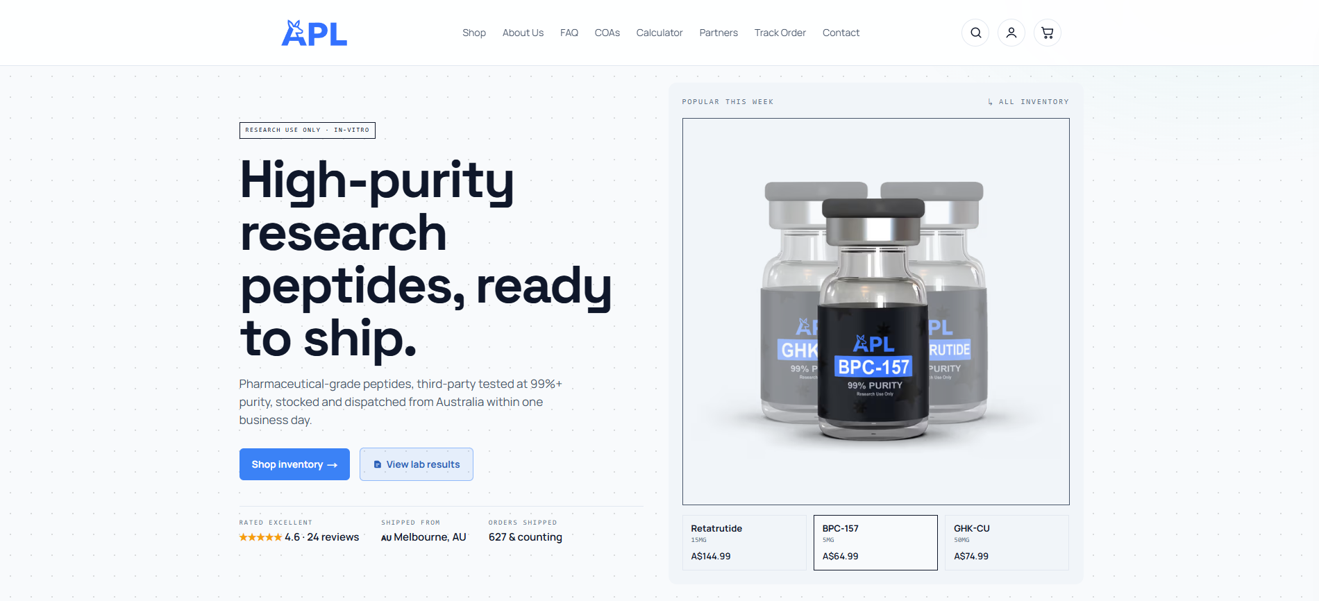
For solubility preparation work, the Peptide Dilution Calculator handles the concentration and volume arithmetic for preparing working stocks from a known lyophilised mass, removing a common source of calculation error. Technical resources on the site cover CoA interpretation, net peptide content, and formulation guidance relevant to the dissolution protocols described in this article. All products are supplied strictly for laboratory research use.
Frequently asked questions
What is the first thing to check when a peptide won't dissolve? Calculate the net charge at the intended assay pH and compare it with the pI. If the pH is within one unit of the pI, shift it by at least two units using dilute acid or base before retrying. If the sequence contains a hydrophobic patch of four or more consecutive nonpolar residues, switch to DMSO or HFIP as the primary solvent.
How do I dissolve a hydrophobic peptide? Dissolve in a small volume of neat DMSO (for Cys/Met-free sequences) or HFIP first, then dilute dropwise into aqueous buffer. Sonication and gentle warming to 37 °C assist dissolution.
What does TFA counter-ion do to solubility? TFA ion-pairs with basic residues (Arg, Lys) and reduces their effective positive charge, which can push a peptide closer to its pI and promote aggregation. Requesting an acetate or HCl salt form from the supplier, or performing a counter-ion exchange by dissolving in ammonium bicarbonate and lyophilising, often rescues the peptide.
How accurate are peptide solubility calculators? Tools like PepCalc provide a useful first-pass solvent-class recommendation based on net charge and pI. CamSol-PTM reports Pearson correlations of approximately 0.58–0.81 between predicted and experimental relative solubility. Both tools predict intrinsic solubility in water, not absolute solubility in a specific assay buffer. Always validate with a small experimental check in the actual assay conditions.
How should I store a dissolved peptide? Store short-term at 4 °C in sealed, low-binding tubes. For longer storage, aliquot into single-use volumes and freeze at −20 °C or −80 °C. Label each aliquot with concentration, solvent, pH and date. Avoid freeze–thaw cycling. Sequences containing Cys, Met or Asn are best stored lyophilised at −80 °C.
When should I use HPLC to measure peptide concentration? HPLC is the gold standard when absolute concentration accuracy is required, when the peptide lacks UV-active residues (no Trp or Tyr), or when degradation or aggregation is suspected. For routine checks, UV absorbance at 280 nm is adequate for Trp/Tyr-containing sequences.
Sources
- Sequence-based prediction of the intrinsic solubility of peptides containing non-natural amino acids | Nature Communications
- Peptide solubility limits: backbone and side-chain interactions (PMC article)
- Measurement and modelling solubility of amino acids and peptides in aqueous 2‑propanol solutions (RSC)
- Peptide solubility calculator
- Peptide solubility prediction: visualising hydrophobicity vs charge (Peptide Chemistry)