
Peptide labelling standards fall into three functional categories, and the correct choice depends entirely on your readout. Fluorescent and chromogenic tags suit detection and kinetic assays, isotopic (heavy) peptides serve as internal standards for mass spectrometry quantification, and affinity tags exist to simplify purification. For FRET-based protease assays, the fluorophore/quencher pairs with the strongest track record are EDANS/Dabcyl and Mca/Dnp, with FAM/TAMRA reserved for applications needing longer wavelengths or multiplexing. Whichever category you select, two non-negotiables apply: verify the final product by RP-HPLC and mass spectrometry, and label the vial with enough detail to trust it six months from now.
Before committing synthesis time or budget, match the category to the experimental goal:
- Fluorescent/chromogenic tags — best for real-time kinetic assays, cellular imaging, and FRET-based protease substrates.
- Isotopic (heavy) standards — the only sound choice for absolute quantification in SRM/MRM mass spectrometry workflows.
- Affinity/purification tags — chosen when isolating labelled product from truncated synthesis by-products matters more than detection sensitivity.
Get the category wrong and you will spend weeks troubleshooting an assay that was never going to work with that tag in the first place.
What are the main categories of peptide labelling standards?
Fluorescent and chromogenic labelling attaches a light-emitting or colour-producing group to a peptide backbone, either at the N-terminus, C-terminus, or a side chain, so the molecule can be tracked by fluorescence, absorbance, or FRET. It is the workhorse category for enzyme kinetics, receptor-binding assays, and cell imaging, because signal generation is immediate and instrument access (a plate reader or fluorimeter) is common in most laboratories. Isotopic labelling replaces one or more atoms with a stable heavy isotope (commonly ¹³C, ¹⁵N, or deuterium) so the resulting peptide behaves chemically identically to its native counterpart but reads as a distinct mass in a spectrometer. Affinity tags, meanwhile, are short sequences or chemical handles (His-tags, biotin, IMAC-compatible motifs) added purely to make purification or immobilisation easier.
Choosing between these three comes down to three practical questions.
- What is the assay readout? If you need a signal a plate reader can capture in real time, fluorescent labelling wins. If you need to distinguish a spiked internal standard from the endogenous peptide in a complex biological matrix, only an isotopic label will do that reliably, because chemically it is indistinguishable from the native sequence.
- Do you need absolute or relative quantification? Fluorescence intensity is useful for relative comparisons within a single assay but is a poor substitute for absolute quantitation, which requires a heavy isotope standard spiked at a known concentration before extraction.
- Does the downstream step involve purification or immobilisation? If you are isolating a labelled peptide from a complex synthesis mixture, an affinity tag such as a His-tag or biotin handle earns its place, even though it adds bulk.
The trade-offs matter as much as the category itself. Fluorescent dyes such as TAMRA or fluorescein are bulky relative to many peptide backbones, and placing a large, planar aromatic group near a binding interface can distort receptor affinity or block an enzyme's active site. Peptide-tag researchers generally recommend using the smallest tag that still accomplishes the experiment, because no single labelling method is perfect: enzymatic tags offer high specificity but slower kinetics, metal-recognition tags label quickly but can suffer from background metal chelation, and peptide-coil interaction strategies (such as E/K coiled-coil pairs) hit a useful middle ground of speed and specificity at a small size.
Isotopic labelling avoids the steric interference problem entirely, since a heavy isotope adds negligible mass and zero chemical bulk, but it comes with its own constraint: you need mass spectrometry access and a reference standard with a documented enrichment level to make the numbers meaningful. Affinity tags sit at the opposite extreme. They are often the bulkiest addition of the three, and biotin or His-tag sequences can interfere with folding or activity if placed carelessly, so chemoselective purification tags are usually cleaved or positioned away from any functional domain once their purification job is done. Toxicity and biocompatibility rarely factor into in vitro assay work, but they become relevant fast if the labelled peptide is destined for any cell-based or in vivo research application.
Selecting fluorophores and quenchers: spectral table and pairing guidance
Getting fluorophore selection wrong is the single most common reason a FRET assay produces flat, uninterpretable data. The fix starts with matching excitation and emission wavelengths to your instrument's filter set and avoiding spectral overlap with assay autofluorescence, which is usually strongest below 450 nm.
The EDANS/Dabcyl pairing is the most cited combination in protease substrate design because Dabcyl's absorbance maximum sits almost exactly on top of EDANS's emission peak, giving efficient quenching at rest and a large fluorescence jump once the enzyme cleaves the peptide between donor and acceptor. Mca/Dnp is the second workhorse pair, favoured when a smaller donor is needed or when the assay wavelength range must stay below 400 nm to avoid interference from other assay components. FAM and TAMRA are less commonly used as a FRET pair on their own (their spectral overlap is weaker than EDANS/Dabcyl), but FAM's brightness and TAMRA's red-shifted emission make each valuable individually for direct fluorescence detection, HPLC tracking, or multiplex imaging where two distinguishable colours are needed on separate peptides.
Beyond the raw spectral numbers, three practical considerations decide whether a labelled peptide performs as expected:
- Isomer purity matters more than most researchers expect. Fluorescein-based dyes like FAM exist as multiple positional isomers (5' vs 6' carboxyfluorescein, for instance), and a mixed-isomer preparation gives inconsistent quantum yield between batches. Insist on isomerically pure starting material.
- FAM's phenolic hydroxyl groups need protection during synthesis. Left unprotected, they can react during repeated piperidine deprotection cycles in Fmoc SPPS, generating side products that show up as extra HPLC peaks. Blocking the phenol by tritylation before continuing the synthesis avoids this entirely.
- Terminal versus side-chain placement changes assay geometry. N- or C-terminal labelling is simpler synthetically and usually preferred for FRET substrates, since it maximises the physical separation between donor and quencher across the cleavage site. Side-chain labelling (via a Lys or Cys residue) is reserved for cases where the terminus is needed for another purpose, such as immobilisation or a second tag.
Choose the pairing before you choose the coupling chemistry. Redesigning a substrate after synthesis because the fluorophore emission overlaps your plate reader's excitation filter wastes far more time than the extra half-hour spent checking a spectral table up front.
Step-by-step fluorescent labelling protocols for SPPS
Solid-phase peptide synthesis gives you two practical routes to a fluorescently labelled peptide: build the dye in during chain assembly, or couple it on after the sequence is complete. Each has a clear use case, and mixing them up wastes resin and reagent.
- Choose a preloaded resin when the label sits at the C-terminus. NovaTag-style preloaded resins arrive with the fluorophore, quencher, or affinity handle already anchored, cutting several synthetic steps compared to a post-synthetic coupling and reducing the risk of incomplete labelling on the final residue. This is the fastest path for standard FRET substrates built around Dansyl, Mca, EDANS, or Dnp.
- Incorporate Fmoc-Lys(dye)-OH directly into the sequence when the label must sit internally. Treat the dye-modified lysine as a standard Fmoc amino acid during chain assembly, but extend the coupling time, since the bulky aromatic side chain slows resin penetration and can leave truncated sequences if coupling is rushed.
- Reserve N-terminal or side-chain post-synthetic coupling for OSu ester dyes. FAM and TAMRA are frequently supplied as N-hydroxysuccinimide (OSu) esters, which react cleanly with a free amine (the N-terminus or a Lys side chain) once the peptide is otherwise complete and still resin-bound or in solution.
- Select the coupling reagent to match the dye's reactivity. PyBOP with DIPEA as base gives fast, reliable amide coupling for most fluorophore carboxylic acids. HOBt/HOAt-mediated activation is gentler and reduces racemisation risk on sensitive residues, while DIPCDI (diisopropylcarbodiimide) paired with HOBt is a solid default for standard dye conjugation where cost matters at scale.
- Extend coupling times for bulky dyes. TAMRA and similar rhodamine-based dyes are sterically demanding; a standard 30 to 60 minute coupling window often under-couples. Extending to 2 to 4 hours, or repeating the coupling once, closes the gap and avoids carrying deletion peptides into purification.
- Protect reactive phenolic groups before piperidine treatment. As noted for FAM, unprotected phenolic hydroxyls react during Fmoc deprotection cycles. Tritylation (Trt-Cl with DIPEA) before further chain extension prevents this.
- Cleave and deprotect under standard TFA cocktail conditions, then check the crude product by analytical HPLC before committing to full-scale purification. A crude trace with a dominant single peak at the expected retention time saves you from purifying a batch that was never going to hit purity targets.
The most common failure mode in fluorescent SPPS is not a chemistry problem, it is a scheduling one: researchers treat the dye-coupling step like any other amino acid addition and move on too quickly. Bulky aromatic dyes need more time on resin, not less, and skipping that step shows up later as a shoulder peak on the HPLC trace that is expensive to chase down after the fact.
Pro Tip: Run a small-scale test coupling (10 to 25 mg resin) with a new dye lot before committing your full synthesis batch. Dye-OSu esters vary in reactivity between suppliers and even between lots, and a quick test coupling followed by a Kaiser or chloranil test for free amine catches a sluggish reagent before it costs you a week of resin time.
Isotopic labelling for mass spectrometry standards
Isotopic peptide standards exist for one purpose: giving mass spectrometry a chemically identical but mass-distinguishable reference so quantification is absolute, not relative. Two supply routes get you there, and the choice depends on how much control you need over the labelling position.
The first route builds the heavy label directly into synthesis using isotopically enriched Fmoc amino acids, ¹³C, ¹⁵N, or deuterium-substituted at a specific residue, so the exact position of the mass shift is known and controllable. The second route sources a full-length heavy peptide as a ready-made internal standard, spiked into a sample at a known concentration before extraction or digestion to correct for losses throughout sample preparation. Stable isotope-enriched amino acids and synthetic heavy peptides are now standard tools in targeted proteomics, particularly for SRM and MRM workflows where absolute quantitation depends entirely on the accuracy of the spiked standard.
Isotopic purity and labelling site both affect how trustworthy your quantification ends up being.
- Isotopic enrichment below about 98% introduces a measurable light-isotope contaminant peak, which skews the ratio calculation between heavy standard and native analyte, particularly at low analyte concentrations where the light peak is proportionally more significant.
- Labelling site selection determines fragmentation behaviour in MS/MS. A heavy label placed on a residue that survives common fragmentation pathways (avoiding proline-adjacent bonds, for instance) gives cleaner, more interpretable spectra.
- The number of labelled atoms per peptide sets your mass shift resolution. A single-atom shift can sit too close to natural isotope envelopes of the light peptide on lower-resolution instruments; multi-atom labelling (a 6 to 10 Da shift) is the safer default for most quadrupole and QTRAP platforms.
On the supply side, always request a certificate of analysis that states isotopic enrichment per labelled atom explicitly, not just as an aggregate purity figure, and treat the MS spectrum on that COA as your acceptance test for incoming stock rather than taking the supplier's purity claim at face value. Storage and shipping matter here too: heavy peptides are chemically identical to native peptides and degrade the same way, so they need the same cold-chain handling, desiccated storage, and light protection as any other peptide standard, regardless of the impressive mass tag sitting on the label.
Purification and quality control for labelled peptides
A labelled peptide is only as good as its purity, and purity is only meaningful once it has been verified independently by more than one method. Chemoselective purification is the first line of defence when a synthesis run contains truncated or deletion sequences alongside the full-length labelled product.
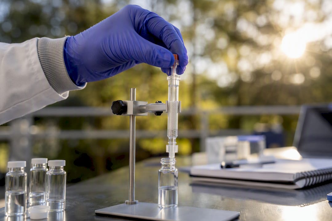
IMAC-based purification uses a metal-loaded IDA (iminodiacetic acid) column, typically running at pH 6.5 to 8.0 for binding and eluting the target peptide around pH 3.5, and works well when the peptide carries a His-tag or comparable metal-binding motif. Biotin-tagged peptides take a different route, captured on avidin or streptavidin resin and eluted either by competitive displacement or, for cleavable linkers, by chemical or enzymatic release. Both chemoselective approaches are worth the extra step when the crude synthesis mixture is complex, since they separate on a property the truncated by-products almost never share.
RP-HPLC remains the workhorse polishing step regardless of whether chemoselective capture was used first.
Mass spec verification is where you confirm the label actually attached where and how you intended.
- Check the observed mass against the calculated mass including the label's contribution, not just the unlabelled peptide backbone; a mismatch of even a few Daltons flags incomplete labelling or an unexpected side reaction.
- Inspect the isotopic envelope shape, since a distorted or split envelope often indicates a mixture of labelled and unlabelled species that HPLC alone failed to separate.
- Document HPLC purity percentage and isolated yield for every batch, not as a formality but as the working record you will need to explain any inconsistency between batches months later.
Falling short of that threshold does not necessarily make the batch useless, but it does mean any quantitative result built on it carries a wider error margin than the assay's other variables probably justify. For a closer look at acceptance criteria and spectral interpretation, the mass spec peptide verification guide walks through the checks in more detail.
Product labelling and vial labelling standards for traceability
A peptide standard with a poorly labelled vial is a liability the moment it leaves the freezer, because six months from now nobody, including you, will remember which lot it came from or when it was reconstituted. Traceability starts with the label, not the freezer log.
A compliant research-use vial should carry five elements at minimum:
- Product name and full sequence (or an unambiguous reference code tied to a documented sequence on file).
- Net quantity, stated in mass or nanomoles, matching the certificate of analysis exactly.
- Lot number, unique to that synthesis and purification batch.
- Expiry or retest date, set conservatively for labelled peptides, which can be more photolabile or hydrolytically sensitive than their unlabelled counterparts.
- A clear "Research Use Only" disclaimer, stated unambiguously and not buried in fine print.
Label formatting matters more than researchers usually credit it for: sans-serif fonts print more legibly at small sizes, a minimum critical font size of 6 to 8 pt keeps text readable on small vials, and high-contrast text (black on white, not coloured text on a coloured background) holds up better under the low lighting common in cold rooms and freezer walk-ins.
Material choice matters just as much as the text itself. Standard adhesive labels lift or smear after repeated freeze-thaw cycles and condensation exposure, so thermal-transfer printed labels or purpose-made cryogenic label stock hold up far better across a standard storage lifecycle. Details on packaging materials suited to cold-chain conditions are covered in the peptide packaging materials guide.
Pro Tip: Print the lot number as both human-readable text and a scannable barcode or QR code where your inventory system supports it. It costs nothing extra at the printing stage and eliminates the transcription errors that creep in when someone hand-copies a lot number into a lab notebook at 6pm.
Record-keeping should tie every vial back to its COA by lot number in a way that survives a staff turnover or a shared-drive reorganisation, not just in the memory of the person who ordered it.
How Au Peptide Labs supports compliant peptide labelling standards
Meeting the standards above depends on the raw material being right before a single coupling reaction happens.
Local dispatch matters more to labelling workflows than it first appears. Every order ships from Australia within one business day, in discreet packaging, cutting out the customs delays that can leave a time-sensitive labelling project stalled at a border while a coupling window closes.
- Purity documentation on request supports the HPLC and mass spec checks covered above, and the net peptide content glossary entry clarifies exactly what that percentage figure represents on a COA.
- Custom and bulk orders are available for researchers who need a specific labelled or isotopically enriched peptide sequence built to order.
- Documentation to request when commissioning custom work: a COA stating both chemical purity and, for isotopic standards, enrichment per labelled atom, along with the MS spectrum used to verify it.
Browse the current research peptide range to see COA and labelling practices applied at the product level, including examples such as the LL-37 and SS-31 listings.
Quick troubleshooting checklist for common labelling failures
Most labelling failures trace back to one of four causes, and diagnosing which one is at fault takes minutes if you know where to look first.
- Low coupling yield on the dye residue. Check the HPLC trace for a truncated peak lacking the dye's expected mass shift. The fix is usually a longer coupling time (2 to 4 hours instead of 30 to 60 minutes for bulky dyes like TAMRA) or a double coupling with fresh activated reagent.
- High background fluorescence in the assay. Rule out unreacted free dye first, since even trace unconjugated fluorophore left over from an incomplete purification inflates baseline signal. A second HPLC polish, or a longer desalting step before the assay, usually resolves it.
- Weak or absent FRET signal despite a correctly labelled substrate. Confirm the donor and quencher are on opposite sides of the intended cleavage site and that the spectral pairing genuinely overlaps; a substrate that reads correctly by mass spec can still fail functionally if the geometry places donor and quencher too far apart at rest.
- Degraded signal or shifted retention time on a standard that worked previously. Check storage conditions first: repeated freeze-thaw cycles, light exposure, or a lapsed expiry date are the usual culprits. If the standard has been in active use for more than a few months past its retest date, request a fresh COA or a batch retest rather than trusting an ageing aliquot.
When two or more of these symptoms appear together, particularly a mass spec result that looks fine alongside a functional assay that does not, the problem is usually geometric (labelling position or tag size interfering with the assay) rather than chemical. No amount of re-purification fixes a design issue.
Storage conditions and stability considerations
Labelled peptides degrade faster than most researchers expect, and the labelling chemistry itself is often the weak point. Store fluorescently labelled peptides desiccated, protected from light, and at minus 20°C or colder for long-term storage; working aliquots can typically tolerate 2 to 8°C for short periods, but repeated freeze-thaw cycling is the single most common cause of gradual signal loss in FRET substrates.
Photobleaching is a real concern for fluorophores like FAM and TAMRA, which lose quantum yield under prolonged light exposure, even ordinary room lighting during a long assay session. Amber vials or foil wrapping during storage and handling reduce this risk substantially.
Isotopically labelled peptides carry the same chemical stability profile as their unlabelled counterparts, since the heavy isotope changes mass, not reactivity, so they need identical storage discipline: desiccated, cold, and light-protected where the sequence contains photosensitive residues such as tryptophan or methionine.
Practically, labs label vials with the compound name, total mass, reconstitution volume, reconstitution date, and a break-in-use date, since a reconstituted peptide standard has a materially shorter working life than the lyophilised powder it came from. Thermal-transfer or thermal-print vial wraps hold up better than standard adhesive labels through repeated cold storage and condensation cycles, keeping that information legible months after the vial first went into the freezer.
Before relying on an older standard for a critical experiment, run a quick HPLC check against the original COA trace. A shifted retention time or a new shoulder peak is the clearest early sign of degradation, well before it shows up as inconsistent assay results.
Calibration protocols for quantitative assays
A labelling standard only earns the name "standard" once it has been used to build a proper calibration curve, not just spiked into a single sample and trusted on faith. For mass spectrometry work, this means preparing a dilution series of the heavy isotope standard across a concentration range that brackets the expected analyte concentration in real samples, then plotting the heavy-to-light peak area ratio against known concentration to establish linearity.
A calibration curve with an R² below roughly 0.99 across the working range signals a problem worth chasing before running real samples, whether that is a degraded standard, an unstable injection, or a concentration range extending beyond the assay's true linear response.
For fluorescence-based assays, calibration works differently but the discipline is the same: run a dilution series of the free fluorophore (not the intact substrate) to establish the maximum theoretical signal at complete cleavage, then use that ceiling value to calculate percentage conversion in kinetic experiments rather than reporting raw fluorescence units, which vary between instruments and even between sessions on the same instrument.
Re-run the calibration curve whenever a new lot of standard comes into use, not just at assay validation. Lot-to-lot variation in isotopic enrichment or fluorophore conjugation efficiency is common enough that treating an old calibration curve as permanent is one of the more avoidable sources of quantitative error in peptide-based assays.
Safety considerations for labelled peptides
Labelled peptides carry the standard handling precautions of any peptide reagent, plus a few specific to the label itself. Fluorescent dyes such as FAM, TAMRA, and Dabcyl are chemically reactive compounds in their free (unconjugated) form, and should be weighed and handled in a fume hood with standard PPE: nitrile gloves, lab coat, and eye protection, given their potential for skin and eye irritation.
Dnp-containing reagents warrant particular care. Dinitrophenyl compounds are sensitising agents in some individuals, and repeated skin contact should be avoided, making glove changes between handling steps a sensible default rather than an optional precaution.
Isotopically labelled peptides using stable isotopes such as ¹³C, ¹⁵N, or deuterium carry no radioactivity and no additional handling risk beyond the peptide itself; they should not be confused with radiolabelled compounds, which fall under an entirely different regulatory and safety framework.
All peptide labelling standards covered in this guide are supplied and intended strictly for laboratory research use, not for human or veterinary administration. Standard laboratory chemical hygiene practice, including working in a certified fume hood for powder handling and proper disposal of dye-containing waste through your institution's chemical waste stream, applies across every category discussed here, regardless of how routine the reagent may seem after repeated use.
Selection criteria based on experimental application
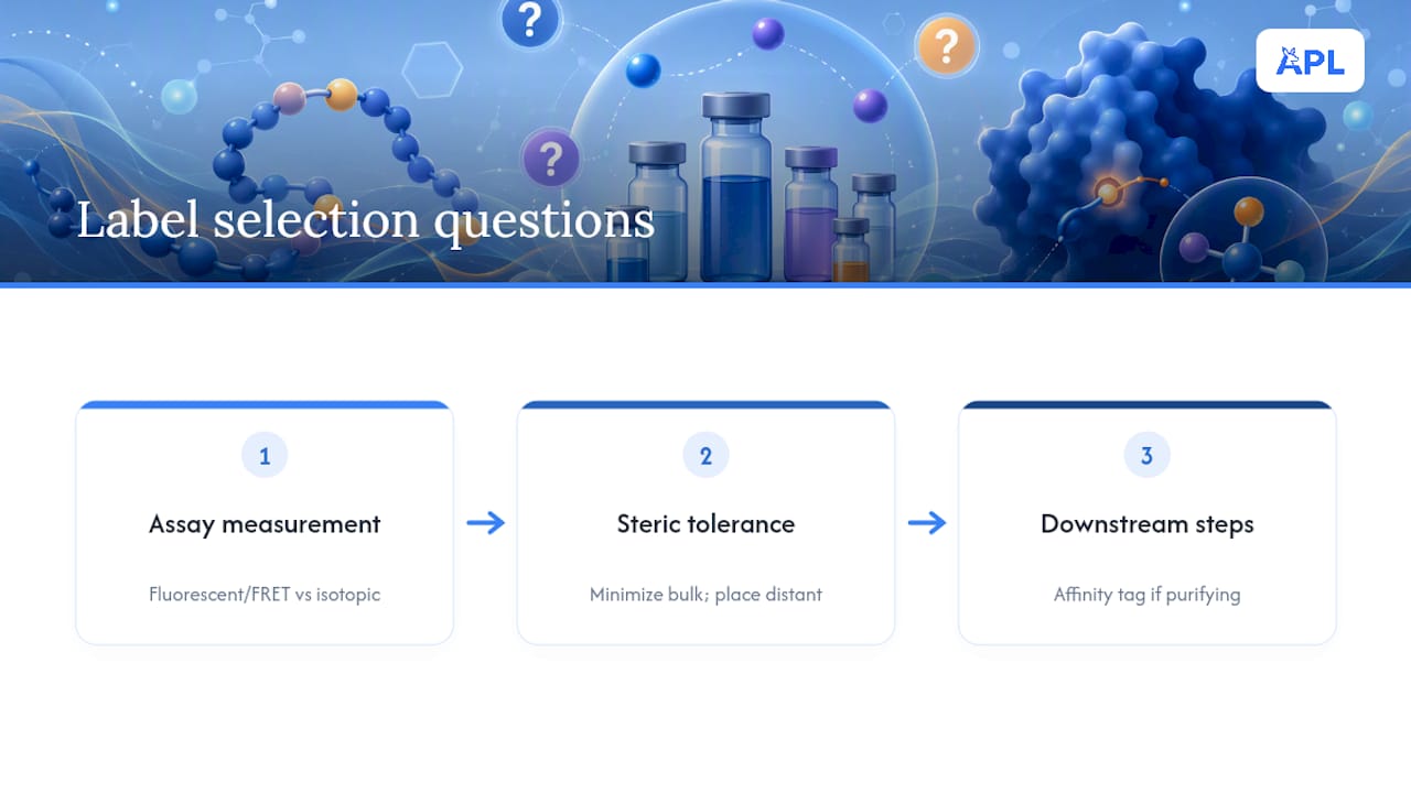
Matching a labelling standard to its experiment comes down to answering three questions in sequence, and skipping any one of them is how researchers end up redesigning a substrate mid-project.
First, what does the assay actually measure? A real-time kinetic readout on a plate reader calls for a fluorescent or FRET-based tag; a quantification question involving absolute concentration in a complex matrix calls for an isotopic standard, full stop, since no fluorescent method delivers that kind of quantitative rigour reliably.
Second, how much steric tolerance does the system have? Enzyme active sites, receptor-binding pockets, and protein-protein interaction surfaces vary enormously in how much bulk they tolerate near the functional region. When in doubt, favour the smallest tag that still gives an interpretable signal, and place it as far from the functional domain as the experimental design allows.
Third, what downstream steps follow labelling? If purification from a complex mixture is required, an affinity tag earns its place even at the cost of added bulk. If the peptide goes straight into an assay without further isolation, that same tag is dead weight, adding synthesis cost and steric risk for no functional return.
Cost and turnaround time are legitimate factors too, but they should be the last consideration, not the first. A cheaper, faster-to-synthesise tag that compromises assay sensitivity or quantitative accuracy costs far more in repeated experiments than the modest premium of getting the selection right the first time.
Editorial perspective: what actually separates a usable standard from a wasted synthesis run
Most guidance on peptide labelling treats spectral tables and coupling chemistry as the hard part, and traceability as an afterthought. That ordering is backwards. The chemistry described in this piece, FRET pairing, SPPS coupling, HPLC polishing, is well understood and rarely the actual point of failure in a working lab. The recurring failure is a mislabelled or under-documented vial six months after synthesis, when nobody can say with confidence which lot, which isotopic enrichment, or which retest date applies to the standard sitting in the freezer.
The conventional advice gets the fluorophore selection and coupling protocol right and then treats the label itself as packaging. It is not packaging. It is the only thing standing between a defensible quantitative result and a guess. Prioritise the boring parts first: request the COA that states isotopic enrichment per atom, not just an aggregate purity number, and insist on a legible, durable label before worrying about which dye pairing looks most elegant on paper. The elegant substrate that nobody can trace back to its batch record is not a standard at all.
— Dr. Authur
Sources
- Peptide-tags for site-specific protein labelling in vitro and in vivo - Molecular BioSystems
- Fluorescence-quenched peptide substrates and protease assays (PMC review)